 |
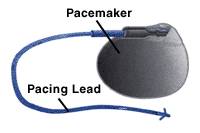
Single-Chamber Pacemakers. In a single-chamber pacemaker, only one wire (pacing lead) is placed into a chamber of the heart. Sometimes it is the upper chamber, or atrium. It also may be placed in a lower chamber, or ventricle. |
|
 Dual-Chamber Pacemakers In dual-chamber pacemakers, wires are placed in two chambers of the heart. One lead paces the atrium and one paces the ventricle. This more closely matches the natural pacing of the heart. Also, this pacemaker can follow the heartbeats originating in the atrial (upper) heart chamber and pace the ventricular (lower) heart chamber to maintain coordinated function between the atria and ventricles. Dual-Chamber Pacemakers In dual-chamber pacemakers, wires are placed in two chambers of the heart. One lead paces the atrium and one paces the ventricle. This more closely matches the natural pacing of the heart. Also, this pacemaker can follow the heartbeats originating in the atrial (upper) heart chamber and pace the ventricular (lower) heart chamber to maintain coordinated function between the atria and ventricles. |
| In the largest clinical trial of pacemakers conducted in the U.S., researchers found that patients with Sick Sinus Syndrome benefit from dual chamber pacemakers. They are less likely to develop atrial fibrillation or mild forms of heart failure compared to people who are treated with single-chamber pacemakers. |
|
|
|
|
How a Pacemaker Works
|
- The pacemaker sends a tiny electrical signal called a "pacing pulse".
- The signal travels through insulated wires, or leads, until it reaches a metal electrode at the tip of the lead.
- The electrode delivers the electrical signal directly to the heart.
- The signal causes the heart tissue to begin the muscle contractions that cause a heartbeat.
|
|
|
|
|
|
|
|
Conditions Treated with Pacemakers
|
|
| Pacemakers may be prescribed for a number of conditions, including: |
|
|
Bradycardia, a condition in which the heart beats too slowly, causing symptoms such as fatigue, dizziness or fainting spells. Bradycardia may be caused by the wear and tear of age, or to conditions such as sick sinus syndrome (SSS), which affects the sinus node (also called sinoatrial node), the heart's natural pacemaker.
Heart block, a condition affecting the atrioventricular (AV) node (electrical connecter between the upper and lower chambers of the heart), a second natural pacemaker in the heart, may also require pacemaker therapy.
[more on Bradycardia] |
 |
Atrial fibrillation (A Fib) is a common heart rhythm disorder (arrhythmia) in which the upper chambers of the heart beat rapidly and chaotically. In a small subset of patients, particularly those with sick sinus syndrome, pacemakers may be helpful to prevent the condition. |
|
|
Heart failure, (HF) a condition in which the heartbeat is not sufficient to supply a normal volume of blood and oxygen to the brain and other parts of the body. The pacemaker is carefully programmed to increase the force of muscle contractions in the heart. This is called "biventricular pacing" or "resynchronization" therapy. |
|
|
 Syncope, a condition best known as the "common faint," is usually not serious. It may not require treatment or, in some cases, may be treated with medications. Some patients faint when their heart rhythm becomes very slow. A small percentage of people have more severe and more frequent fainting spells. This problem can cause injury, and can impact on a patient's everyday activities. They may be unable to perform their jobs or drive a vehicle for fear they will faint suddenly and cause injury to themselves or others.
[more on syncope] Syncope, a condition best known as the "common faint," is usually not serious. It may not require treatment or, in some cases, may be treated with medications. Some patients faint when their heart rhythm becomes very slow. A small percentage of people have more severe and more frequent fainting spells. This problem can cause injury, and can impact on a patient's everyday activities. They may be unable to perform their jobs or drive a vehicle for fear they will faint suddenly and cause injury to themselves or others.
[more on syncope]
| When a person faints, from neurocardiogenic or vagal syncope the brain sends an inappropriate signal to the heart to slow the heart rate and lower blood pressure. In selected patients, a pacemaker can prevent the heart rate from becoming too slow and avoid a fainting episode. |
|
|
|
ICDs Also Can be Pacemakers
An implantable cardioverter defibrillator (ICD) is a pacemaker-like device used to treat people who are at high risk for ventricular fibrillation (VF), a deadly arrhythmia that kills more than 400,000 people each year and is the number one cause of sudden cardiac death. In VF, the heart suddenly and without warning begins to beat very rapidly and out of rhythm. The heart in VF cannot pump blood, and death occurs within minutes unless a device called a defibrillator is available to deliver an electric shock that restores the heart's normal rhythm. An ICD is a built-in defibrillator. It continuously monitors the heartbeat and automatically shocks the heart back to normal rhythm if it detects VF or tachycardia - a dangerously rapid heartbeat.
ICDs also can function as pacemakers to maintain the heart's normal rhythm after an episode of VF, or when the patient's heart rate is sometimes too fast and sometimes too slow. |
|
| Many people with pacemakers also take medications. Certain medications can affect the proper function of a pacemaker, or affect the interaction between the pacemaker and the underlying heart rhythm disorder. Pacemaker patients should make sure that their heart rhythm specialist is aware of all medications they are taking.This includes over-the-counter medications, diet supplements and herbal products. Many substances available without a prescription are known to affect the rhythm of the heart. In some patients, these products can be life threatening. |
|
|
|
|
|
|
|
The Heart's Natural Pacemakers
|
|
|
|
The sinoatrial (SA) node (also called the sinus node) is the heart's master pacemaker. In the normal heart, the SA node sends electrical signals in a steady, rhythmic pattern to pace the heart's beat. Signals from the SA node travel to: |
|
|
The atrioventricular (AV node), which transmits the signal from the upper chambers, or atria, to the lower chambers (ventricles). The ventricles are the major pumping chambers of the heart. The electrical signal transmitted from the SA and AV nodes trigger the muscle contractions that pump blood out of the ventricles and into the lungs and body. |
|
|

 A pacemaker is a small, electronic device that regulates, or paces, the rhythm of the heart. It usually is the first line of treatment for bradycardia (a heart rate that is too slow).