top-banner
Heart Failure Tests

doctor and patient, color photo
The diagnosis of HF usually begins with a complete physical examination. The doctor also will take a medical history and ask about symptoms. Blood tests probably will be ordered to assess kidney and liver function, levels of sodium and potassium, or other factors that point to a diagnosis of heart failure.
Table of Contents
Chest X-ray
Electrocardiogram (ECG)
Ejection Fraction (EF)
Echocardiography (Echo)
Radionuclide Ventriculography
Electrophysiology (EP) Study
Heart Failure Classifications
The first heart tests that generally are ordered by the physician are:
A chest X-ray, to see if the heart is enlarged or if there is excess fluid in the heart or lungs, and
ECG, color illustration An electrocardiogram (ECG) An ECG is a simple, painless test that often is performed in the doctor's office. The ECG prints out a graph that shows how the heart is beating and records electrical activity of the heart that may point to structural abnormalities.
If these tests suggest a diagnosis of heart failure, the next step may be to measure the heart's ejection fraction (EF). Based on the patient's medical history and symptoms, the doctor will order one or both of the following tests to measure the EF and diagnose whether the problem is systolic failure (the muscle of the lower chamber, or ventricle, loses some ability to pump blood out of the heart) or diastolic failure (the muscles of the hearts' chambers becomes stiff and cannot relax to receive the normal volume of blood):
Ejection Fraction is a measure of the proportion (or fraction) of blood that is pushed out by the ventricle with each contraction, or heart beat. A normal heart pumps out one-half to two-thirds of the blood in the left ventricle with each heartbeat. If the ejection fraction falls below 55 percent, it is a sign that the heart muscle is weakened.
Echocardiography (Echo) is a safe, painless test that uses sound waves (ultrasound) to examine the heart's structure and motion. The patient lies still as a technician moves a device called a transducer over the chest. The transducer gives off silent sound waves that bounce off the heart. The sound waves create moving images of the chambers and valves of the heart that are viewed on a video monitor. This test provides information about how the heart is pumping and how blood is flowing in the heart and blood vessels.    
It shows how large the heart is and how the heart valves are working. It also can estimate the pressure in the heart and determine if there is fluid around the heart.
An Echo creates moving pictures of the heart.
Radionuclide ventriculography (also called the "first pass" technique or Multiple-Gated Acquisition Scanning - MUGA) is a type of nuclear medicine test. A small amount of radioactive dye is injected into a vein. Isotopes in the dye attach to red cells in the blood, and a special camera is used to trace them as they pass through the heart and into the circulation. The efficiency with which the isotopes are pumped out of the heart and travel through the body is a measure of the heart's pumping ability. The dye used for this test is only mildly radioactive and is not harmful. Its radioactivity is short-lived and the dye is excreted in the urine. Sometimes, the images created by the camera are synchronized with an electrocardiogram (ECG) that records the electrical activity of the heart at the same time. A nuclear medicine scan may be given twice: once when the patient is at rest, and again when he or she is exercising. This can show whether a heart that works fairly well at rest has trouble keeping up with the stress of physical exertion.
treadmill exercise, color photo
Treadmill exercise test measures the heart's exercise capacity and how much oxygen the heart provides to muscles during exercise.
Electrophysiology (EP) Study. In an EP study, local anesthetics are used to numb areas in the groin or near the neck, and small catheters are passed into the heart to record its electrical signals. During the study, the physician studies the speed and flow of electrical impulses through the heart, identifies rhythm problems and pinpoints areas in the heart's muscle that give rise to abnormal electrical signals. The doctor uses information from these various heart tests to determine the type and severity of heart failure and what the short-term and long-term outlook (prognosis) may be. They also are important in deciding which treatments are likely to be most effective.
 
An EP study is an electrical "test drive" to determine if an individual's heart has the tendency to go into dangerous rhythms that require treatment.

Heart Failure Classifications

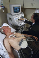
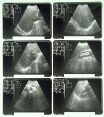
After the physical exam, blood and heart tests have been completed and a diagnosis of HF is confirmed, the physician usually will classify, or "rank," the patient's heart failure. The ranking is based on how severe the symptoms are. The most commonly used system to classify heart failure is called the New York Heart Association (NYHA) Functional Classification. Heart failure patients are placed in one of four categories, depending on how their condition affects the performance of normal physical activities. The four categories are: patient and caregiver, color photoClass I: People in this category feel no symptoms and can perform ordinary physical activity without any limitations. Approximately 35 percent of people with heart failure are in Class I. Class II: Another 35 percent of people with heart failure are in Class II. They have mild symptoms, such as occasional swelling (edema), and may be somewhat limited in their ability to exercise or do other strenuous activities. They do not feel any symptoms when they are at rest. Class III: These individuals are noticeably limited in their ability to exercise or participate in mildly strenuous activities. They are comfortable only when they are at rest. About 25 percent of people with heart failure are in this class. Class IV: The most severe form of heart failure occurs in about 5 percent of patients. These individuals are severely limited in their ability to perform any activity, and they have symptoms even when they are resting.

Leave a Reply

Your email address will not be published. Required fields are marked *